Mechanical Ventilation: 150+ Visual Maps Band 5 Nurses New Starters in ITU International Nurses Respiratory Physios
Ventilator modes, settings, waveforms and alarms explained in visual maps, for nurses and clinicians new to ICU, critical care and HDU.
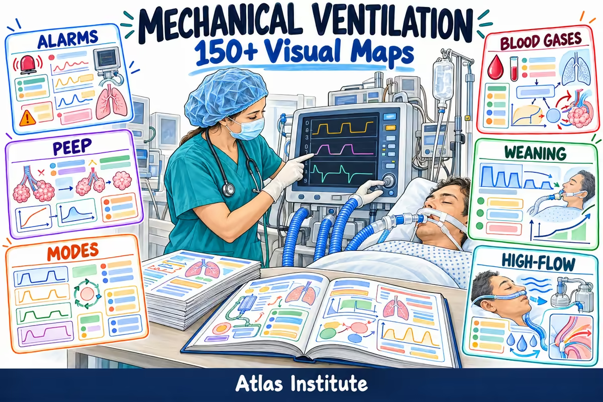
Take a look inside
Real pages from the guide.


The ventilator shouldn’t scare you.
Tick what you’ve been through:
- Hearing the alarm and not knowing if the problem is the patient, the tube or the machine
- Staring at the waveforms without knowing what they’re telling you
- Mixing up PEEP, peak and plateau pressure when you check the settings
- Reading an ABG in kPa and not linking it to the ventilator settings
- Starting on ITU with a few supernumerary shifts and a folder of policies
- Revising ventilation from a 400-page textbook and forgetting it all on your first night shift
This guide is for you.
Ventilation in maps, not in a textbook
Stop dreading the alarms and start reading the ventilator. One topic, one map, and on your next shift you know what’s going on.
The 17 sections of the guide
Click a section to see what’s inside.
10 maps: objectives and indications of mechanical ventilation, the respiratory cycle, airway anatomy, respiratory mechanics, compliance and resistance, ventilation/perfusion (V/Q), gas exchange, classification and diagnosis of acute respiratory failure, managing acute respiratory failure, oxygen delivery devices (nasal cannula, Venturi mask, non-rebreather mask).
10 maps: indications for intubation, the intubation trolley, rapid sequence induction (RSI), the endotracheal tube and cuff, cuff pressure, securing the tube, confirming tube position (waveform capnography, auscultation, chest X-ray), the tracheostomy tube, tracheostomy care, unplanned extubation.
8 maps: ventilator components, assembling the circuit, the pre-use check, HME vs heated humidifier, bacterial filters, closed suction, changing the circuit, the transport ventilator.
12 maps: volume vs pressure, volume control (VC/CMV), pressure control (PC), SIMV, pressure support (PSV), CPAP, dual-control modes (PRVC, volume guarantee), BIPAP and APRV, mandatory vs assisted breaths, trigger, cycling, choosing a mode.
12 maps: tidal volume and predicted body weight, respiratory rate, FiO2, PEEP, inspiratory pressure, peak pressure, plateau pressure, driving pressure, I:E ratio, inspiratory flow, rise time, lung-protective ventilation (6 mL/kg PBW).
6 maps: reading a waveform in 3 steps, pressure-time, flow-time, volume-time, the pressure-volume loop, the flow-volume loop.
7 maps: predicted body weight and tidal volume, static compliance, airway resistance, intrinsic PEEP, capnography (EtCO2), SpO2 targets, haemodynamic monitoring.
7 maps: recognising asynchrony, ineffective effort, double trigger, auto-trigger, flow starvation, premature cycling, delayed cycling.
12 maps: high pressure, low pressure and leaks, low minute volume, high minute volume, apnoea, FiO2, disconnection, tube obstruction, high respiratory rate, PEEP not reached, technical alarms (gas and power), DOPE.
9 maps: sampling and normal values in kPa, pH, PaCO2, bicarbonate, respiratory acidosis, respiratory alkalosis, metabolic acidosis, metabolic alkalosis, the P/F ratio.
11 maps: endotracheal suctioning, mouth care and the VAP bundle, head of bed 30–45°, sedation and RASS, pain (CPOT, BPS), delirium and CAM-ICU, communication, device-related pressure ulcers, enteral feeding, intra-hospital transfer, early mobilisation.
6 maps: initial ventilation in ARDS, indications for proning, proning step by step, care during proning, returning to supine, ECMO referral criteria.
10 maps: readiness to wean, the spontaneous breathing trial (SBT), RSBI, preparing for extubation (cuff leak test), steroids before extubation, the extubation procedure, post-extubation stridor and monitoring, weaning failure, extubation failure, tracheostomy in prolonged weaning.
10 maps: goals and indications of NIV, contraindications, interfaces (mask and hood), the CPAP hood, modes and settings, starting NIV, weaning off NIV, NIV failure and when to intubate, complications, high-flow nasal oxygen (HFNO).
14 maps: ventilator-induced lung injury (VILI), barotrauma, volutrauma, atelectrauma, biotrauma, pneumothorax, ventilator-associated pneumonia (VAP), oxygen toxicity, cardiovascular complications, neurological complications, ICU-acquired weakness, sudden desaturation, COPD on the ventilator, cardiac arrest in the ventilated patient.
6 maps: key differences in children, tubes and cuffs, settings, neonatal ventilation, nasal CPAP, alarms and safety.
8 maps: ARDS from pneumonia, COPD exacerbation on NIV, acute pulmonary oedema on CPAP, the patient fighting the ventilator, desaturation after transfer, intrinsic PEEP in acute asthma, difficult weaning, hypotension after intubation.
Get your ventilation maps
- 150+ visual maps, one topic per map
- Modes, settings, waveforms, asynchronies, alarms, ABGs, NIV, weaning, paediatrics and clinical cases
- Bonus 1: Ventilator Alarms in 60 Seconds
- Bonus 2: ABGs in 3 Questions (in kPa)
- Bonus 3: Shift Checklist for the Ventilated Patient
- Landscape PDF, printable, with space for your notes
- 7-day money-back guarantee

7-day money-back guarantee
If within 7 days you feel the guide hasn’t helped you, we’ll refund 100% of your purchase. No questions asked.
I want the mapsFrequently asked questions
Band 5 nurses new to ICU, ITU or HDU, internationally educated nurses working in UK critical care, respiratory physios, ACCPs in training and resident doctors rotating through critical care.
Yes. It starts from the basics (what the ventilator does, the respiratory cycle, the main settings) and builds up to modes, alarms and waveforms, one map at a time.
No. Each map uses short sentences, labelled drawings and the words you’ll hear on the unit (sats, ABG, PEEP, registrar). The pictures carry the meaning, so it works well if English is your second language.
Yes. Blood gases are in kPa, glucose and lactate in mmol/L, and drugs use their UK names (adrenaline, noradrenaline, salbutamol).
No. It works alongside your unit’s training, local protocols and national guidance (ICS, FICM, NICE). Settings and mode changes are always as prescribed.
It isn’t CPD-accredited. You can record your reading as self-directed learning in your NMC revalidation portfolio, as you would with any study material.
Straight after payment you get an email with access to the members’ area, where you can read the PDF on your phone, tablet or computer, or download and print it.